
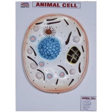
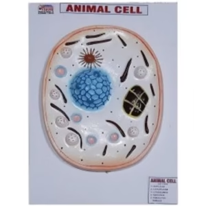
Animal cell models are often used in education to help students understand the complex structure and function of animal cells. They can show the various organelles, such as the nucleus, mitochondria, endoplasmic reticulum, Golgi apparatus, and others, as well as their locations within the cell. These models can provide students with a deeper understanding of the cell and its role in maintaining the health of the organism.
The model visually represents various cell organelles with different colors and textures for educational purposes. The key labeled structures include:
- Nucleus – The control center of the cell, shown in blue.
- Cytoplasm – The gel-like substance filling the cell.
- Lysosomes – Small organelles involved in digestion, depicted as round pink structures.
- Vacuole – A membrane-bound organelle for storage.
- Pinocytic Tubule – Structures involved in fluid intake.
- The model visually represents various cell organelles with different colors and textures for educational purposes.
This model visually represents the internal structure of a Animal cell for educational purposes. We can increase our knowledge and study with the help of this model.


Reviews
There are no reviews yet.